Towards more effective tendon repair with a multi-functional biomaterial
A Janus-faced biomaterial that firmly adheres to injured tendons and helps them glide, also slowly releases drugs to tendon cells, which could improve repair in patients across multiple injuries

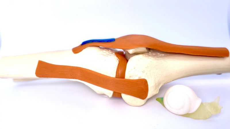
The development of the Janus Tough Adhesive originally was inspired by the tough mucus produced by the Dusky Arion (Arion subfucus) slug. Shown here, it is applied to a tendon within a joint, where its two sides, respectively, enable tight adhesion to tendon tissue and gliding of the protected tendon within its normal joint environment. Credit: Wyss Institute at Harvard University
Many of us have injured a tendon either abruptly during sports activities, or as a result of daily wear-and-tear from overusing them. Torn or strained tendons – most commonly Achilles, flexor, or rotator cuff tendons – can cause persistent pain, and take a long time to heal after surgeries, especially in the elderly. This is not surprising since the brilliant white cords that connect our muscles to our bones have to regenerate their ability to transmit the strong mechanical forces that allow our joints to move.
There is an acute, unmet medical need for more effective therapies to treat tendon injuries. Despite advances in surgical methods, new graft therapies, and more effective rehabilitation, severely injured tendons rarely regain the structural integrity and mechanical strength of healthy tendons, which often places a chronic burden on patients.
Now, a collaborative research team led by David Mooney, the Robert P. Pinkas Family Professor of Bioengineering at the Harvard John A. Paulson School of Engineering and Applied Sciences (SEAS), and Core Faculty member at Harvard’s Wyss Institute for Biologically Inspired Engineering, Eckhard Weber, of the Novartis Institutes for BioMedical Research (NIBR), and Daniel Kaufmann, of the Novartis Technical Research and Development, developed a new biomaterial-based tendon therapy that addresses key challenges in the regeneration process. The two-sided material firmly adheres to tendons with one of its specifically engineered surfaces, while allowing normal gliding of healing tendons with its opposite mechanically tough yet elastic surface. Importantly, “Janus Tough Adhesives” (JTAs) can also act as high-capacity drug depots, slowly releasing small molecules into tendon tissue to help facilitate healing. The researchers compellingly demonstrated the regenerative abilities of drug-free and drug-loaded JTA versions in a series of injury-relevant, pre-clinical tendon models, and reported their findings in Nature Biomedical Engineering.
“JTAs with their combination of tissue-specific capabilities offer a new opportunity to overcome current insufficiencies in tendon regeneration across multiple types of injuries, and could help many patients regain more normal tendon functions and mobility,” said Mooney. “Our exceptional partnership with multi-disciplinary research groups at Novartis, besides providing highly complementary technical, modeling and experimental expertise, also enabled us, for the first time, to optimize and apply a Tough Gel Adhesive for the local delivery of a small molecule drug, which was one of our early goals.”
JTAs are the latest example of biomaterial-based adhesives developed by Mooney’s team as part of their Tough Gel Adhesive platform technology, which originally was inspired by the tough mucus produced by the Dusky Arion (Arion subfuscus) slug. Previously, the researchers had engineered non-toxic, hydrogel-based biomaterials that can adhere to diverse wet surfaces, including injured tissues still covered with blood, and withstand the mechanical stress produced by friction in the moving tissues. These features make them highly useful as tissue sealants (adhesives used to hold tissues together that were severed by injuries) and wound dressings, as current adhesive materials don’t fulfill these essential requirements.
To create Tough Gel Adhesives tailored to tendon regeneration, “we forward-engineered an alginate-polyacrylamide-based hydrogel into a two-sided biomaterial, essentially, by modifying one of its surfaces with a sugar known as chitosan, which we showed allows firm bonding to tendons. The unmodified side could simultaneously enable gliding under the continued friction tendons that are exposed during movement,” said first-author and Postdoctoral Fellow Benjamin Freedman, who spearheaded the study in Mooney’s lab in close collaboration with the Novartis research groups. “Within this ‘Janus-faced’ hydrogel, we then loaded a model drug that could be slowly released and chemically reduce inflammation.”
Janus Tough Adhesives for Tendon Repair from Wyss Institute on Vimeo.
Freedman and his co-workers are developing JTAs as off-the-shelf biomaterials for easy use by surgeons. All the biomaterial components in JTAs, including chitosan, which is obtained from the shells of crustaceans, are biocompatible and already used in other medical applications.
At the start of the project, Freedman had a mini sabbatical at Novartis in Basel, Switzerland, to collaboratively define and stress-test the targeted profile of the tendon therapeutic. A vivid back-and-forth of experimental results obtained on both sides of the Atlantic, and monthly conference calls between the Harvard and the Novartis teams propelled the research forward. In their concerted effort, the teams demonstrated that JTAs robustly bonded to blood-coated isolated porcine tendons and they found them to bind stronger to the different types of tendons than the existing tissue adhesives TISSEEL, Dermabond, and SurgiCel. They also implanted JTAs next to the patellar, foot flexor, and Achilles tendons of a rat in vivo model and confirmed their strong tendon adhesion non-invasively with magnetic resonance imaging (MRI) and high frequency ultrasound.
To show that JTAs’ resilience and elasticity protected tendons and enabled them to glide, the researchers attached them to the flexor tendons of human cadaveric hands, extending and flexing the wrists over hundreds of cycles: they noticed no signs of wear or disintegration.
“Importantly, when we applied JTAs to ruptured patellar tendons of rats, they remained in place over their three-week implantation and facilitated tendon healing. They also reduced the formation of scars by 25%, compared to surgically repaired tendons that we didn’t treat with JTAs,” said Freedman.
Scarring normally accompanies healing as a result of inflammation and causes long-term loss of tendon functionality.
Using a patellar injury model in rats, the researchers investigated the effects of a corticosteroid, an anti-inflammatory model drug, that was encapsulated and released in a sustained manner from JTAs. Using state-of-the-art imaging technologies and molecular immunoassays, they showed that, in the tendons treated with corticosteroid-loaded JTAs, the inflammation subsided significantly faster.
“Therapies for tendon repair and regeneration should be ideally adaptive by nature, and cover the spectrum from resolution of inflammation and pain to restoration of tendon tissue matrix and function,” said Michaela Kneissel, Global Head of the Musculoskeletal Disease Area at NIBR. “The combination of active pharmaceutical ingredients with Janus Tough Adhesives has the potential to become a promising avenue for such an adaptive therapy, with release and activity of pharmaceutical ingredients gated by the JTAs to better facilitate tendon repair and regeneration,” she added.
Other authors on the study are Sungmin Nam, Daniel Kent, Anna Rock, and Yann Tinguely on Mooney’s team at Harvard; Markus Kurz, Andreas Kuttler, Nicolau Beckmann, Michael Schuleit, Farshad Ramazani, Nathalie Accart, Andreas Fisch, and Thomas Ullrich, members of the research teams at Novartis; and Jianyu Li, Assistant Professor at McGill University in Montreal, Canada, who co-developed the first Tough Gel Adhesives in Mooney’s group.